A low-cost 3D cell culture system for imaging neurons
05-04-2018
Current neuronal cell culture is mostly performed on two-dimensional (2D) surfaces, which lack many of the important features of the native environment of neurons, including topographical cues, deformable extracellular matrix, and spatial isotropy or anisotropy in three dimensions. Although three-dimensional (3D) cell culture systems provide a more physiologically relevant environment than 2D systems, their popularity is greatly hampered by the lack of easy-to-make-and-use devices.
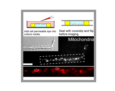
Yuan Ren, a graduate student in the Suter lab has recently developed a simple microwell device for 3D neuronal cell culture that is inexpensive, easy to assemble, and fully compatible with commonly used imaging techniques, including super-resolution microscopy, which was accomplished in collaboration with the group of Dr. Fang Huang in the Weldon School of Biomedical Engineering. Using this device, the researchers found that the morphology and growth pattern of bag cell growth cones in 3D culture closely resemble the ones of growth cones observed in vivo. This microwell device will hopefully facilitate a wider adoption of 3D neuronal cultures to study the mechanisms of neurite regeneration.
A low-cost microwell device for high-resolution imaging of neurite outgrowth in 3D (2018). Yuan Ren, Michael J Mlodzianoski, Aih Cheun Lee, Fang Huang and Daniel M Suter, Journal of Neural Engineering, Volume 15, Number 3.